Take the example of a concerning growth on a patient’s neck, which has doctors considering surgery. There are risks: operating on the neck, where several arteries are located, can be dangerous. Plus, doctors aren’t completely sure the growth is cancerous, so maybe the procedure isn’t necessary.
In the future, thanks to a new type of imaging scanner being developed by researchers and students at the Universities of Canterbury and Otago (with help from Christchurch Polytechnic Institute of Technology), medical professionals should be able to determine if such surgeries are necessary – before they’re performed.
Known as the Medipix All Resolution System (MARS), the imaging machine is able to take multi-energy “true colour” X-ray images and analyse what is shown in the picture, such as the interior contents of a tumour.
Behind the MARS is the father-son duo of UC professor Dr Phil Butler and associate professor Dr Anthony Butler. In addition, at least 20 current or completed UC PhD students have been involved over the last few years, along with masters and honours students.
Phil Butler says the MARS represents a significant improvement over traditional X-ray or magnetic resonance imaging (MRI), which can only analyse the shapes of objects and not the content that makes them up.
“If you’ve got an infection in a knee joint replacement, with a standard system you can’t actually see the infection,” he says. “But you’ll be able to see it with this.”
Part of the secret to the technology lies with a 14mm-square chip, a semiconductor that measures the particle properties of an X-ray photon, as well as its energy and position.
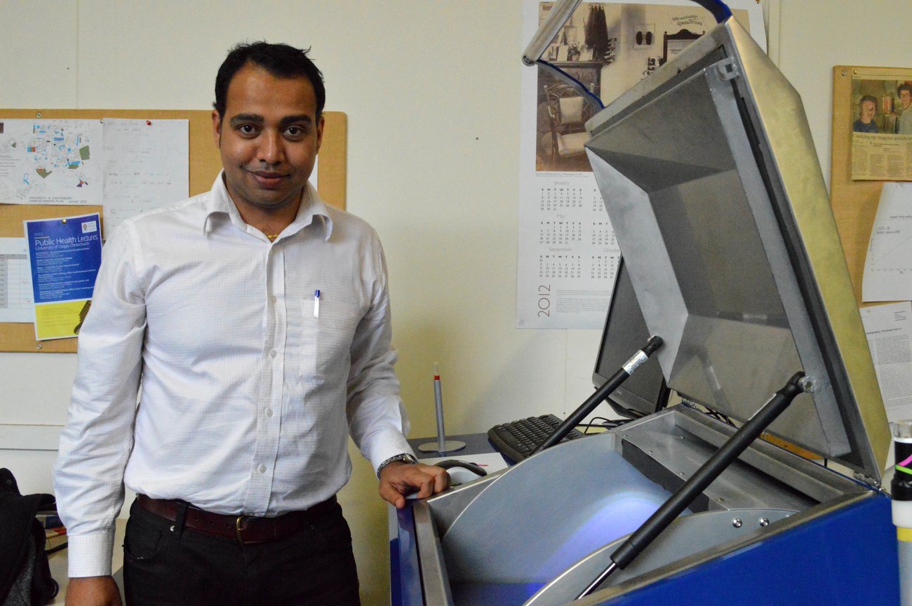
A tissue sample or small animal (a mouse, for example) can be placed inside the MARS, which takes a few minutes to create 3D images.
The images are then reconstructed and recorded on a computer, so they can then be accessed and analysed.

“You can identify or characterise or measure components of a tissue [with the MARS],” says Anthony Butler. “You can measure the water content or the fat content or the calcium content [for example]. If you compare that to an ordinary X-ray, you can’t do that at all.”
Funding for the MARS began in 2002 with a $350,000 grant from the New Economy Research Fund.
A $1.5 million grant from the Tertiary Education Fund followed later, as did an additional $4.5 million from what is now known as the Ministry of Business, Innovation and Employment (MBIE), which went towards developing a small animal version.
In late 2014, the MBIE awarded $12 million for research into a human-sized version of the MARS – meaning the scanner has received more than $20 million in direct Government funding over more than a decade.
The Butlers point out that the funding figures don’t include university support through student scholarships, staff time and access to equipment. University-sourced funding is comparable to that provided by Government, Phil Butler says.
While the Butlers acknowledge there are many applications for the MARS, their work has focused on medicine.
“We’ve had to work more closely with the medical researchers who have brought their own skill sets,” says Anthony Butler.
“So we’ve been working with people who look at blood vessels or atheromas [when degenerative material accumulates in artery walls] or vascular disease, and particularly around strokes and the blood vessels in the neck. We’ve been working with cancer researchers, and we’ve been working a lot with people on joint implants and metal implants for knee replacements and cartilage health. But there are dozens of other applications.”
As a result of research, the MARS project has spawned a commercialisation spin-off company, Mars Bioimaging Ltd (MBI). Founded by the Butlers in 2007, the company has sold six MARS devices for about $500,000 each, with two going to the US, three to Europe, and one staying in New Zealand.
A human-sized version of the MARS is expected to be built by – or possibly before – 2020, Butler says.
The father-son team are also founding members of the Medipix-3 collaboration, which has seen 24 institutions across the globe join forces on X-ray imaging research.
In addition, researchers from institutions including Yale and the Mayo Clinic have collaborated on the MARS, while other researchers and scientists regularly visit.
Domestically, there are plans to use the MARS to look at brain disease in sheep, in collaboration with Lincoln University.
Dr Nigel Johnson, the University of Canterbury’s director of Research & Innovation, says there are now exciting opportunities to develop and manufacture key componentry for human scanners.
“It is taking a large research team from numerous institutions to pull this off, and we are fortunate to have had strong support from New Zealand government agencies.”